Submitted by Emily Rigby on Mon, 14/07/2025 - 12:03
A new paper from the Herbison lab, published in Nature Communications, investigates how stress has a substantial negative impact on fertility.
Noradrenergic (AN) neurons are considered to be one group of cells that convey stress signals within the brain. A new study led by Dr Szilvia Vas and Dr Paul Morris in the Herbison laboratory at the University of Cambridge has utilized an array of advanced ex vivo and in vivo approaches in the mouse to reveal a direct mechanism through which heightened activity in brainstem NA neurons can pause pulsatile reproductive hormone secretion in a sex- and gonadal steroid-dependent manner.
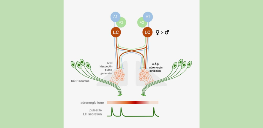
Using brain slice electrophysiology, the scientists demonstrated that arcuate nucleus kisspeptin (ARNKISS) neurons, recently identified to be the gonadotropin-releasing hormone (GnRH) pulse generator, are directly hyperpolarized by NA through both alpha 2- and beta-adrenergic receptors. Retrograde viral tracing showed that NA innervation of the ARN is primarily from the dorsal subdivision of the locus coeruleus (LC)-NA cell group and is substantially greater in females compared to males. Using an intersectional genetic approach allowing selective chemogenetic manipulation of NA neurons innervating the ARN alongside photometry recording of ARNKISS neuron synchronization behaviour, they find that the activation of NA inputs strongly suppresses GnRH pulse generator activity in a sexually differentiated and gonadal steroid-dependent manner. These results indicate a likely pathway through which excessive stress would suppress the brain mechanisms that drive reproduction. Read the full paper, Brainstem noradrenergic modulation of the kisspeptin neuron GnRH pulse generator in mice.